변이 우간동맥에 의한 총담관 압박으로 발생한 폐쇄황달 1예
Obstructive Jaundice Due to Compression of the Common Bile Duct by Variant Right Hepatic Artery
Article information
Abstract
간담도계 동맥에 의해 총담관이 압박된 예는 매우 드물다. 우간동맥에 의해 총담관이 압박되어 폐쇄황달이 발생하였을 경우, 수술적 감압으로 확진 및 치료를 할 수 있다. 20세 남자 환자가 황달을 주소로 내원하였다. 전산화 단층촬영 혈관 조영술에서 위십이지장 동맥에서 분지한 변이 우간동맥에 의한 총담관 압박으로 발생한 폐쇄황달 소견을 보였다. 내시경 경비담관 배액술을 시행하였고, 수일 후 복강경 담낭절제술, 복강경 변이 우간동맥 가동술과 T자관 삽입술 시행하였다. 환자는 별다른 급성기 합병증은 없었고, 이후 1년간 재발도 없었다.
Trans Abstract
Extrahepatic bile duct can be compressed by right hepatic artery (RHA) and cause a variety of hepatobiliary symptoms. This condition is referred to as RHA syndrome. A 20-year-old man was admitted because of jaundice. No stones or tumor were visible on CT scan and endoscopic retrograde cholangiopancreatography. However, RHA was seen traversing and compressing the mid common bile duct (CBD) with resultant upstream dilatation. The patient was diagnosed with obstructive jaundice due to compression of the CBD by variant RHA originating from gastroduodenal artery. After separation and mobilization of the variant RHA, obstructive jaundice was resolved. Herein, we report a case of a variant form of RHA syndrome that was successfully managed by surgery.
서 론
폐쇄황달은 총담관결석증, 종양, 췌장염, 수술이나 시술 후 담도 협착, 기생충 감염, 간담도계 혈관의 담관압박 등에 의해 발생할 수 있다. 간담도계 혈관에 의해 총담관이 압박된 예는 드문데, 우간동맥, 위십이지장 동맥에서 분지한 우간동맥, 총간동맥, 췌십이지장동맥 가성동맥류등에 의한 총담관 압박이 보고된 바 있다[1-7]. 이 중 간담도계에 변이 혈관이 존재하고 이 혈관에 의해 폐쇄 황달을 일으키는 경우는 더욱 드물다. 저자들은 황달을 주소로 내원한 20세 남자 환자에게서 위십이지장 동맥에서 분지한 변이 우간동맥에 의한 총담관 압박으로 발생한 폐쇄황달을 진단하였다. 내시경 경비담관 배액술을 통해 배액을 시행하였으며 수술적 감압을 통해 성공적으로 치료한 증례를 경험하였기에 보고하는 바이다.
증 례
특이 과거 질환이 없는 20세 남자가 내원 수 일 전부터 발생한 황달을 주소로 내원하였다. 전신적 신체검사상 혈압은 110/70 mmHg, 맥박수 72 회/분, 체온 36.7oC이었고 만성 병색 소견과 황달 결막 소견을 보였으며 복부 진찰에서 압통은 없었다. 말초 혈액 검사에서 백혈구 7,850/mm3, 혈색소 12.5 g/dL, 혈소판 337,000/mm3 이었다. 혈액 화학 검사에서 총 빌리루빈 14.4 mg/dL, 직접빌리루빈 10.4 mg/dL, ALP 2,061 IU/L, AST 207 IU/L, ALT 296 IU/L, 총 단백 6.9 g/dL, 알부민 4.1 g/dL, 혈중 요소 질소 11.4 mg/dL, 크레아티닌 1.0 mg/dL 이었다. 응고 검사에서 프로트롬빈시간은 10.7초(INR: 1.04)로 측정되었다.
복부 전산화 단층촬영을 시행하였고 총담관 중간부위에 협착과 그 근위부에 담관 확장이 있었으나 담관 내부에 담관석이나 종양은 없었다. 그리고 협착 부위를 가로지르는 변이 동맥이 관찰되었다(Fig. 1A). 내시경 역행 췌담관 조영술(endoscopic retrograde cholangiopancreatography, ERCP)에서 바터 팽대부에 특이 소견은 없었고, 담관 조영술에서는 복부 전산화 단층촬영과 동일한 부위인 총담관 중간부위에 밴드형의 조영제 충만 결손과 협착이 관찰되었다(Fig. 1B).
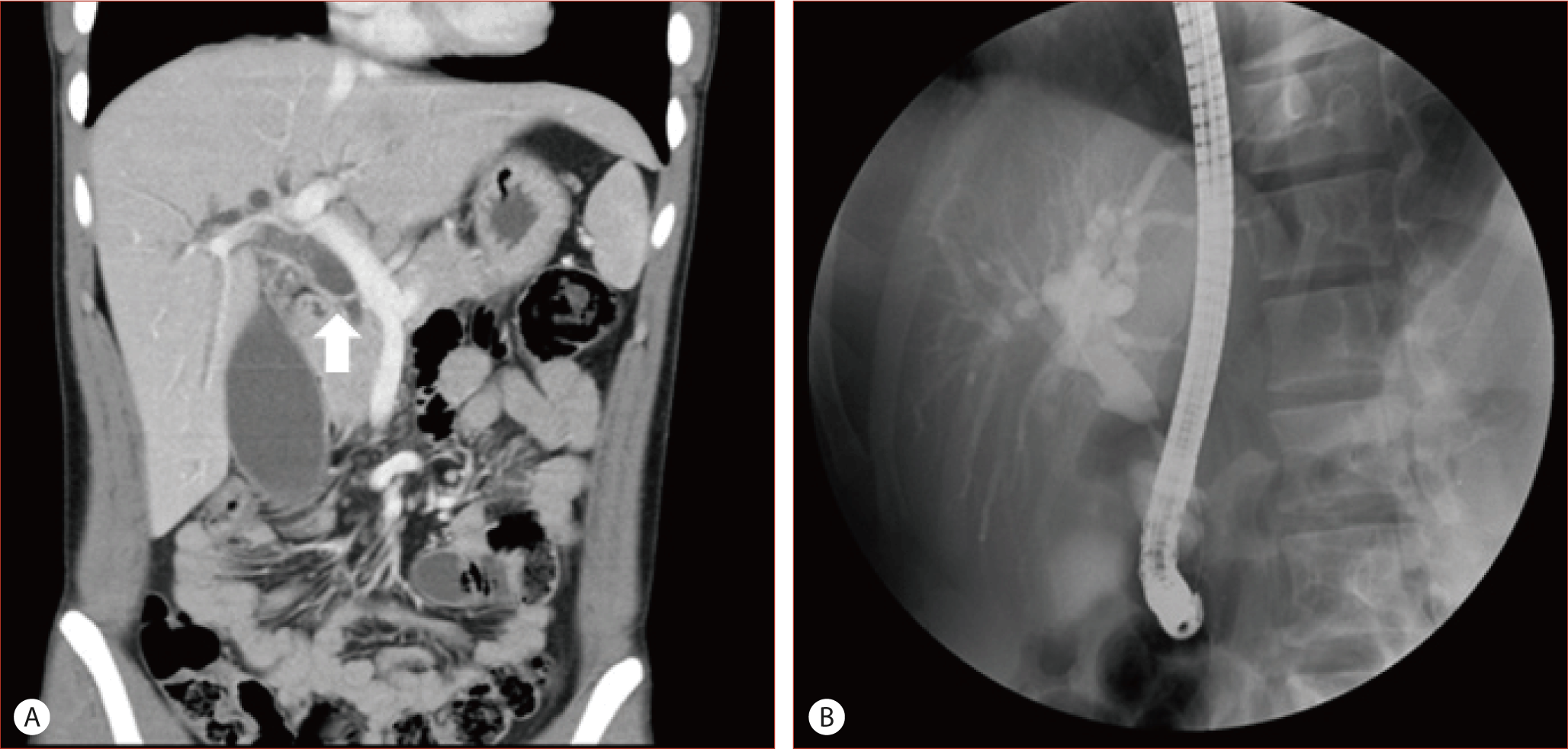
(A) Abdominal CT image shows proximal bile duct dilatation upstream to the point where hepatic artery traverses the common bile duct (CBD) (arrow). (B) Endoscopic retrograde cholangiopancreatography shows a band like filling defect at the mid CBD.
담즙의 배액을 위해 내시경 경비담관 배액술(endoscopic nasobiliary drainage, ENBD)을 시행하였고 이후 환자의 총 빌리루빈, 직접빌리루빈, ALP, AST, ALT가 감소하였다. 동맥과 담관의 상관관계를 확인하기 위해 ENBD 카테터를 유지한 채로 전산화 단층촬영 혈관 조영술(CT angiography)을 시행하였다. 3차원으로 재구성한 CT angiography 영상에서 고유간동맥 외에 위십이지장 동맥에서 분지한 변이 우간동맥이 추가로 존재하였고, 변이 우간동맥에 의해 중간부위 총 담관이 압박되는 것이 관찰되었다(Fig. 2).
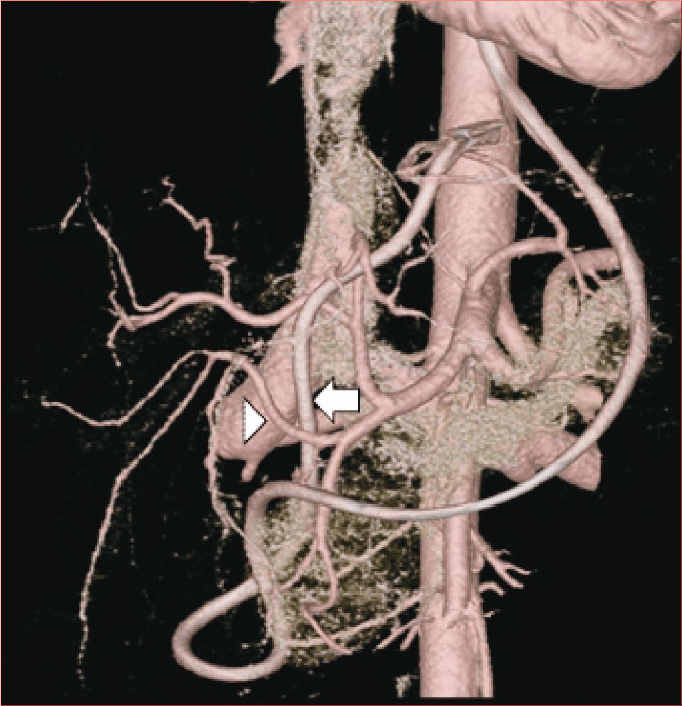
Reconstructed CT angiography image shows a variant right hepatic artery (arrow head) originating from gastroduodenal artery and transversing the common bile duct at the mid portion. Endoscopic nasobiliary drainage catheter (white arrow) is observed in this image.
환자 근본적인 치료 및 확진을 위해서 수술을 시행하였다. 수술 중에서도 CT 소견과 동일하게 위십이지장 동맥에서 분지한 변이 우간동맥이 총담관 앞쪽으로 지나며 총담관을 압박하는 것이 확인되었다. 감압을 위해서 복강경 변이 우간동맥 가동술(laparoscopic mobilization of the variant right hepatic artery)을 시행하였고, 복강경 담낭 절제술과 T자관 삽입술(laparoscopic cholecystectomy & T-tube choledochostomy)도 함께 시행하였다(Fig. 3). 수술과 관련된 합병증은 없었고 환자는 수술 후 5일째 퇴원하였다. 수술 한 달 후 시행한 T자관 담관 조영술에서 총담관 중간부위의 협착은 더 이상 관찰되지 않았으며, 담관 확장도 호전된 소견을 보였다(Fig. 4). 재협착 방지를 위해 T자관은 7주간 유지 후 제거되었으며 한 달간 외래 추적검사에서 총 빌리루빈, 직접빌리루빈, ALP, AST, ALT는 모두 정상화되었고, 이 후 일 년 동안 재발은 관찰되지 않았다.
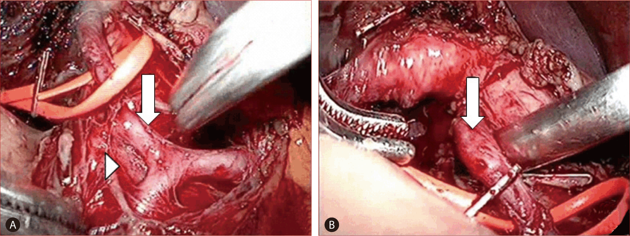
Operative findings. Variant right hepatic artery (white arrow) crossing anteriorly and compressing the common bile duct (arrow head) is observed.

T-tube cholangiography performed one month after surgery. The stenotic lesion at the mid common bile duct (CBD) has disappeared and the proximal CBD dilatation has also improved.
고 찰
간담도계 혈관에 의한 담관 압박으로 발생한 폐쇄황달 증례는 매우 드물게 보고되고 있다. 그중에서 우간동맥에 의한 간외 담관 압박으로 인해 폐쇄황달이 발생하면 우간동맥 증후군이라 한다[5]. 간담도계의 혈관 중 간동맥의 해부학적 변이는 비교적 잘 알려져 있는데[8,9], 간이식환자 1,000예의 간동맥의 변형을 연구한 논문에서는 약 75%에서만 정상 해부학적 구조를 보인다고 보고하였다[8]. 그중에서도 위십이지장 동맥에서 분지한 간동맥에 대한 보고는 매우 드물다. Koops 등은 604명에게서 혈관 조영술을 통해 간동맥의 해부학적 변이를 연구하였고, 이 중 단 두 명에서만 위십이장 동맥에서 분지한 좌간동맥의 예가 있었다[9].
동맥 압박에 의한 폐쇄황달의 진단을 위해서 transabdominal ultrasonography, CT, ERCP, percutaneous transhepatic cholangiography (PTC), endoscopic ultrasonography, magnetic resonance cholangiopancreatography, three-dimensional contrast-enhanced magnetic resonance angiography, digital subtraction angiography 등의 다양한 검사 방법들이 시행되고 있다. Multislice helical CT cholangiography를 이용하여 우간동맥에 의한 총담관 압박을 진단한 예도 있었다[10]. 진단을 위해서 담관과 동맥의 상관관계를 보는 것이 중요한데, 본 증례에서는 ENBD 카테터를 유지한 채로 전산화 단층촬영 혈관 조영술을 시행하였고 3차원 재구성 영상을 이용하여 동맥의 총담관 압박을 확인하였다. 좀 더 침습적인 방법으로 혈관 조영술과 T자관을 이용한 담관 조영술을 동시에 시행하여 동맥 압박을 진단한 경우도 있었다[4]. 하지만 확진은 유발 동맥의 감압이라는 치료와 함께 가능하다.
치료는 수술을 통한 원인 동맥의 감압이 대부분이다[1-5,10]. 수술적 치료 전 배액을 위해 percutaneous transhepatic bile drainage, ENBD 등이 시도되고 있으며[1,3,5], 혈관 압박과 함께 담관석이 동반된 경우라면 담관석 제거를 위해 ERCP, PTC 등을 이용한 담관석 제거가 고려될 수는 있겠지만 근본적인 치료는 아니다. 증상이 없이 총담관 확장만 동반되며 혈액학적 검사실 검사에서 특이 소견을 보이지 않는 경우에는 정기적으로 경과를 관찰하는 경우도 있다[6].
우간동맥 증후군에 간담도계 변이가 동반된 경우도 있는데, Chung 등은 간좌엽의 외측 부재가 동반된 우간동맥 증후군 증례를 보고하였고[2], Miyashita 등은 등 쪽에서 간외 담관을 압박하는 우간동맥 증후군을 보고 하였다[5]. 본 증례처럼 우간동맥 증후군과 위십이지장 동맥에서 분지한 간동맥 변이가 동반된 경우도 드물게 보고되고 있다. Baek 등은 위십이지장 동맥에서 분지한 우간동맥의 총담관 압박과 담관석에 의한 폐쇄황달 환자 증례를 보고하였다[1]. 비록 Baek 등이 보고한 증례에서는 우간동맥이 총담관을 압박하며 지나고 있었으나 압박 부위 상방에 담관 확장, 담관석이 존재하여 폐쇄황달의 원인이 단지 혈관 압박에만 기인한다고 보기는 어렵다. 무증상 담관석에 의한 만성 염증으로 담도 협착이 유발되었고 이에 폐쇄황달이 발생하였을 가능성 또는 담관석 자체에 의한 폐쇄황달의 가능성도 있어 보인다. 본 증례에서는 담관석이 존재하지 않았고, 혈관 압박에 의한 외부 압박 외의 다른 폐쇄황달의 원인이 없다는 점에서 위 증례와 차이가 있다.
저자들은 담관석, 종양 등의 담관 내부의 다른 담즙정체 유발 인자 없이 위십이지장 동맥에서 분지한 변이 우간동맥 압박에 의해 발생한 폐쇄황달 증례를 경험하였고 내시경 경비 담관 배액술과 수술적 감압 후 1년간의 추적관찰 동안 합병증, 재발 없이 성공적으로 치료한 증례를 경험하였기에 보고하는 바이다.
Notes
The author has no conflicts to disclose.