췌장암과 감별이 어려웠던 췌장의 방선균증
A Solitary Pancreatic Actinomycosis Mimicking Pancreatic Cancer
Article information
Abstract
방선균증(actinomycosis)은 실 모양의 혐기성 세균인 방선균에 의한 감염 질환으로, 유황과립이 특징적인 화농성 병변을 초래한다. 방선균증은 종괴처럼 보이는 병변으로 발현할 수 있는데, 이런 이유로 방선균증은 종종 악성 종양으로 오인됐고, 많은 환자들이 정확한 진단이 내려지기 전에 수술적 절제를 받았다.
저자들은 만성췌장염 환자에게 발생한, 악성 종양과 감별이 어려웠던 췌장에 국한된 방선균증 증례를 보고하는 바이다. 환자는 감염의 증상 및 징후를 전혀 보이지 않았고, 다른 장기의 동반 감염도 없었다. 모든 영상 소견은 염증성 종괴보다는 췌장암을 더 시사했다. 그는 내시경초음파 유도 가는 바늘 생검을 통해서 수술 없이 방선균증을 진단 받았다. 한 달 동안의 항생제 치료 이후에 췌장의 종괴는 완전히 사라졌다.
Trans Abstract
Actinomycosis is a chronic, slowly progressive, and suppurative disease caused by filamentous anaerobic bacteria Actinomyces, which results in characteristic sulfur granules. Clinically, actinomycosis can present with a mass-like lesion, and this bacterial nidus has been frequently mistaken for a malignancy. For that reason many patients undergo surgical resection before the correct diagnosis is established. We report a case of a 63-year-old man with a solitary, asymptomatic pancreatic actinomycosis that masqueraded as pancreatic cancer. He did not have any other concurrently infected organs and did not have any signs or symptoms of infection. All radiologic images of the patient favored a malignancy to a great extent rather than an inflammatory mass. He was finally diagnosed with actinomycosis by endoscopic ultrasound (EUS)-guided fine needle aspiration biopsy without surgery. After one month of treatment with antibiotics, the pancreatic head mass was completely resolved on the follow-up computed tomography (CT).
INTRODUCTION
Actinomycosis is an uncommon, slowly progressive, and suppurative disease caused by the genus Actinomyces, which are gram-positive, pleomorphic but commonly filamentous, and strict or facultative anaerobic bacteria. Actinomyces are commensal inhabitants of the oral cavity and intestinal tract. When mucosal integrity is broken, the bacteria can invade local organs and become pathogenic [1].
Actinomycosis can occur in a variety of regions; the oral-cervicofacial, thoracic, and abdominopelvic regions are frequently involved. Approximately 15-20% of actinomycoses occur as an intra-abdominal infection. Abdominal actinomycosis can occasionally present with a mass-like lesion, and the infection focus has been frequently mistaken for a malignancy. Unfortunately, among these cases less than 10% are properly diagnosed prior to surgery [2].
We here report a case of solitary pancreatic actinomycosis, which mimics pancreatic cancer in a patient with a history of chronic pancreatitis. In the literature, there have been only a few cases of actinomycosis that involved the pancreas. Most of these cases however showed either other concurrently infected organs or signs of infection, from which an infection could be suspected without difficulty. To our knowledge, out current patient is the first reported case of a solitary, asymptomatic pancreatic actinomycosis that was initially suspected to be a malignancy on radiologic imagery but then accurately diagnosed by endoscopic ultrasound (EUS)-guided fine needle aspiration biopsy without surgery.
CASE REPORT
A 63-year-old man with a medical history of chronic pancreatitis was hospitalized with a pancreatic head mass that was discovered incidentally by computed tomography (CT), which he had checked as part of an annual surveillance for underlying chronic pancreatitis.
PAST MEDICAL HISTORY
Five years previously, the patient was diagnosed with chronic pancreatitis during his regular medical checkup without any symptoms. He did not have any well-known risk factors for chronic pancreatitis such as alcohol intake. Genetic testing was therefore performed for an unexplained chronic pancreatitis. A c.88-1A>G mutation of the SPINK1 (serine protease inhibitor Kazal-type 1) and c.623G>C mutation of the PRSS1 (protease, serine, 1) genes were identified. He was ultimatedly diagnosed with gene-associated chronic pancreatitis. At that time, he was treated with endoscopic pancreatic sphincterotomy due to stricture of the pancreatic duct orifice and upstream pancreatic duct dilatation.
PRESENT ILLNESS
Since the diagnosis of chronic pancreatitis the patient had never presented any symptoms such as fever, abdominal pain, and weight loss. He was hospitalized for the evaluation and management of the pancreatic mass. At this admission, he did not report any complaints.
Laboratory findings
The results of laboratory tests were as follows: a white blood count (WBC) count of 6,900/mm3; aspartate transaminase (AST) level of 12 IU/L and alanine transaminase (ALT) level of 10 IU/L; serum amylase and lipase levels of 58 U/L and 10 U/L, respectively; and a C-reactive protein (CRP) level of 0.1 mg/dL. The initial serum level of carbohydrate antigen 19-9 (CA19-9) was 8.2 U/mL (reference range: 0-37 U/mL).
Radiologic findings
Dynamic computed tomography (CT) of the pancreas revealed diffuse parenchymal atrophy and multiple parenchymal calcifications with an irregular duct dilatation, which suggested chronic pancreatitis. The dilatation of the pancreatic duct seemed more prominent than that of 5 years ago. The CT scan demonstrated a small, low-attenuation mass in the pancreatic head (Fig. 1A). Magnetic resonance cholangiopancreatography (MRCP) depicted this mass as an irregular shaped, T1 low signal intensity lesion with adjacent parenchymal enhancement on a gadolinium-enhanced, fatsuppressed, T1-weighted image and diffusion restriction on a diffusion weighted image (Fig. 1B). F-18 fluorodeoxyglucose (FDG) whole body positron emission tomography (PET) showed a focal hypermetabolic lesion at the corresponding site of the pancreatic head with a maximum standard uptake value (SUVmax) of 4.0 (Fig. 1C). There were no hypermetabolic lesions other than the pancreatic mass. These imaging data indicated a solid parenchymal mass in the pancreas, which was highly suggestive of malignancy.
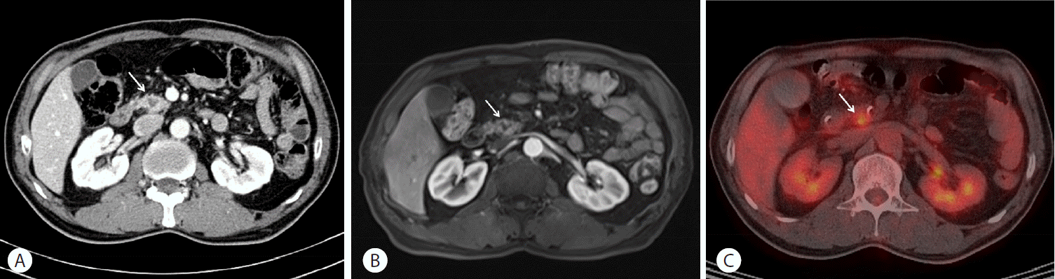
(A) Dynamic computed tomography (CT) of the pancreas demonstrating a low-attenuation lesion (arrow) in the pancreatic head. (B) Magnetic resonance cholangiopancreatography (MRCP) depicting this mass (arrow) as an irregular shaped, T1 low signal intensity lesion with adjacent parenchymal enhancement on a gadolinium-enhanced, fat-suppressed, T1-weighted image. (C) F-18 fluorodeoxyglucose (FDG) whole body positron emission tomography (PET) showing a focal hypermetabolic lesion (arrow) at the corresponding site with a maximum standardized uptake value (SUVmax) of 4.0.
Endoscopic findings
On endoscopic retrograde pancreatography, a focal stricture of the main pancreatic duct at the genu portion and an irregular dilatation of the upstream pancreatic duct with multiple filling defects in it were noted (Fig. 2A). The multiple filling defects, which were identified as protein plugs, were removed by ductal sweeping using a retrieval balloon. EUS revealed a 15-mm sized, ill-defined, hypoechoic mass in the pancreatic head (Fig. 2B). An EUS-guided fine needle aspiration biopsy of that lesion using a 22-gauge needle was performed to exclude malignancy.

(A) A focal stricture (arrowheads) of the main pancreatic duct at the genu portion, an irregular dilatation of the upstream pancreatic duct, and multiple filling defects (arrows) were noted on endoscopic retrograde pancreatography. (B) Endoscopic ultrasound (EUS) revealed a 15-mm sized, illdefined, hypoechoic mass in the pancreatic head. An EUS-guided fine needle aspiration biopsy using a 22-gauge needle was performed.
Histopathological findings
Microscopically, the biopsy specimen of the pancreas revealed active suppurative inflammatory cells surrounding large colonies of filamentous microorganisms, which resulted in characteristic sulfur granules. On hematoxylin and eosin staining multiple microabscesses and large colonies of Actinomyces were shown. Gomori’s methenamin silver stain demonstrated multiple branching filamentous microorganisms or Actinomycetes (Fig. 3A, B).
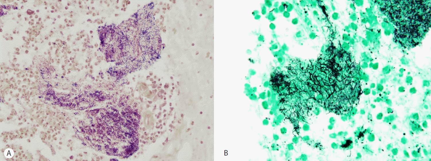
(A) Endoscopic ultrasound (EUS)-guided fine needle aspiration biopsy of the pancreas showing multiple microabscesses and large colonies of Actinomyces, forming characteristic sulfur granules (hematoxylin and eosin stain, ×400). (B) Multiple branching filamentous microorganisms represent Actinomyces (Gomori’s methenamin silver stain, ×1000).
Treatment
The patient was finally diagnosed with pancreatic actinomycosis, which obviated the need for surgery. He was treated with intravenous penicillin G 24 million units per day divided every 4 hours for 2 weeks followed by oral amoxicillin 2,000 mg per day divided every 6 hours for 2 weeks. The pancreatic head mass was completely resolved on the follow-up computed tomography (CT) taken one month after treatment with antibiotics (Fig. 4).
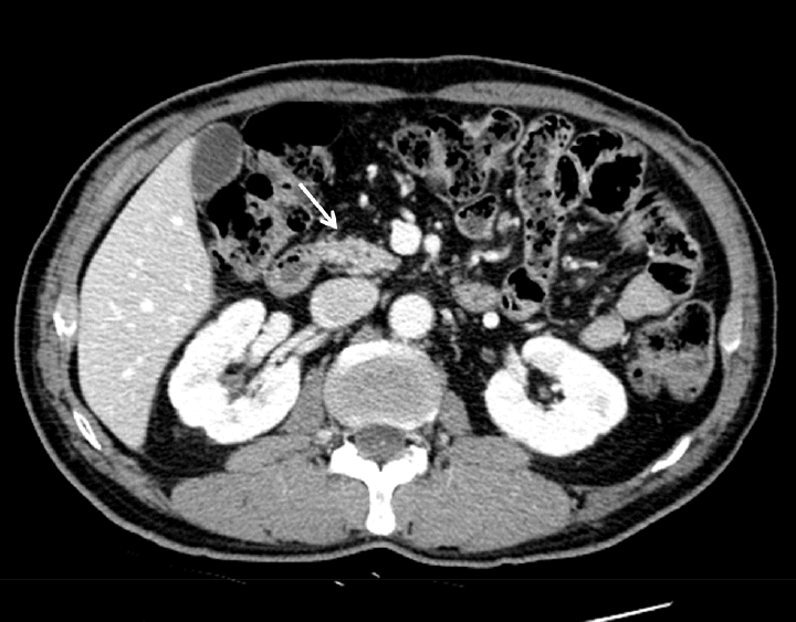
After one month of treatment with antibiotics, the pancreatic head mass was completely resolved on the follow-up computed tomography (CT).
DISCUSSION
Actinomycosis is a rare, chronic infection characterized by abscess formation, tissue fibrosis, draining sinus, and the release of typical sulfur granules. Actinomyces israelli is the most common human pathogen and is found in most clinical presentations of actinomyceal infection. Less common species include A naeslundii, A odontolyticus, A viscosus, A meyeri, and A. gerencseriae [1].
Actinomyceal infection commonly affects three areas. The majority (>50%) involve the oral-cervicofacial region, and disease at this site is associated with poor oral hygiene and dentition. Thoracic actinomycosis represents 15-20% of cases, and disease at this site may present as a diffuse pulmonary infiltration or a discrete mass mimicking bronchial carcinoma. Actinomycosis of the abdomen and pelvis accounts for approximately 20% of cases [2].
Pancreatic actinomycosis is even rarer, and there have been only a few reported cases of actinomycosis involving the pancreas (Table 1). Six of the nine reported pancreatic actinomycosis cases in the literature are male. The median age of patients with pancreatic actinomycosis was 55 years. Most patients presented with nonspecific symptoms such as abdominal pain, loss of appetite, and weight loss, among which the most common symptom was abdominal pain. Five patients had an underlying chronic pancreatitis. Previously reported cases were mostly accompanied by the signs of infection such as fever, leukocytosis, and elevated C-reactive protein (CRP). Other infected organs besides the pancreas were also observed in four of the previous cases (Table 1). The presence of either the signs of infection or other concurrently infected organs would help to differentiate an inflammatory pancreatic mass from malignancy. However, our patient had a solitary pancreatic mass and did not show any signs or symptoms. Therefore, our patient was not thought to be a case caused by infection initially.

Previously reported cases of actinomycosis involving the pancreas
The portal of entry for Actinomyces species is typically a preceding break in the mucosal integrity of the gastrointestinal tract. This infection is frequently linked to bowel perforation, abdominal trauma, intestinal surgery, or intrauterine contraceptive devices [11]. A previous history of ERCP may be a risk factor for pancreatic actinomycosis [9]. However, the patient of our case had no history of endoscopic or surgical pancreatic intervention for the past 5 years prior to this admission. We believe that chronic pancreatitis itself might be the predisposing factor for pancreatic actinomycosis in this patient. There have been several case reports of pancreatic actinomycosis in patients who had a history of chronic pancreatitis similar to our present case [5-7,9]. Progression of underlying chronic pancreatitis may result in structural damage to the parenchyma and ductal epithelium, which could be a potential entry for actinomyceal invasion.
Pancreatic actinomycosis is difficult to diagnose because of its rarity and nonspecific clinical features. Common signs and symptoms include fever, abdominal pain, weight loss, and leukocytosis. In more advanced cases, actinomycosis could be accompanied by extensive sinus, fistula, and abscess formation [8]. Our present patient was thought to have been in an early stage of infection, which is the reason why he did not show any symptoms or signs.
Furthermore, pancreatic actinomycosis can present as a mass-like lesion, and it could be often confused with malignancy. In our present case, the presence of risk factors for pancreatic cancer such as old age and underlying chronic pancreatitis, and the absence of inflammatory signs or symptoms including fever, leukocytosis, or elevated C-reactive protein (CRP) strongly suggested pancreatic cancer rather than an inflammatory mass. Differentiating between an inflammatory mass and a malignancy was possible prior to surgery with an EUS-guided fine needle aspiration biopsy.
The diagnosis of actinomycosis depends on the culture of the pathogen itself or on the histological identification of specific sulfur granules [2]. Sulfur granules represent colonies of Actinomyces and are characterized by a zone of granulation tissue surrounding one or more oval eosinophilic granules. Filamentous gram-positive bacilli radiate from these granules.
The treatment of choice for actinomycosis is high-dose penicillin (18-24 million units per day). For penicillin-allergic patients, tetracycline, erythromycin, or clindamycin are acceptable alternatives [11]. The duration of therapy varies from several weeks to several months based on the initial burden of actinomycosis, but the response is usually favorable.
In conclusion, pancreatic actinomycosis is a rare infection which can masquerade as a malignancy. An accurate preoperative diagnosis is important to minimize morbidity caused by this disease and to avoid unnecessary surgery. An EUS-guided fine needle aspiration biopsy may be helpful to make a preoperative histological diagnosis.
Notes
Conflict of Interest
The author has no conflicts to disclose.