담관낭종 불완전 절제 후 잔류 낭종에 발생한 거대 췌석
Large Pancreatolith Formed in the Remnant Cyst after Incomplete Excision of Choledochal Cyst
Article information
A 44-year-old female was admitted due to recurrent epigastric pain. She had undergone excision for a congenital choledochal cyst at the age of 27 years. Laboratory findings were as follows: white blood cell count of 4,600/mm3, total bilirubin of 0.7 mg/dL, aspartate transaminase of 38 IU/L, alanine transaminase of 20 IU/L, gamma-glutamyl transpeptidase of 30 IU/L, and serum amylase of 242 IU/L. Both abdominal computed tomography and magnetic resonance cholangiopancreatography showed findings compatible with remnant choledochal cyst with 15 mm sized pancreatic stone in the head portion (Fig. 1, 2). Endoscopic retrograde cholangiopancreatography revealed presence of anomalous pancreatobiliary junction, remnant choledochal cyst, and a filling defect in the pancreatic duct (Fig. 3). A white, fragile stone was removed after endoscopic sphincterotomy (Fig. 4). The patient remained asymptomatic without any complications after discharge. Although management of remnant choledochal cyst without symptoms is less clear [1,2], endoscopic sphincterotomy may be useful for the treatment and prevention of pancreatolith in patients with remnant choledochal cyst. Furthermore, complete excision of congenital choledochal cyst is necessary [3]. Incomplete resection of the cyst may cause pancreatic duct stone or protein plug formation in the remnant choledochal cyst [4,5].
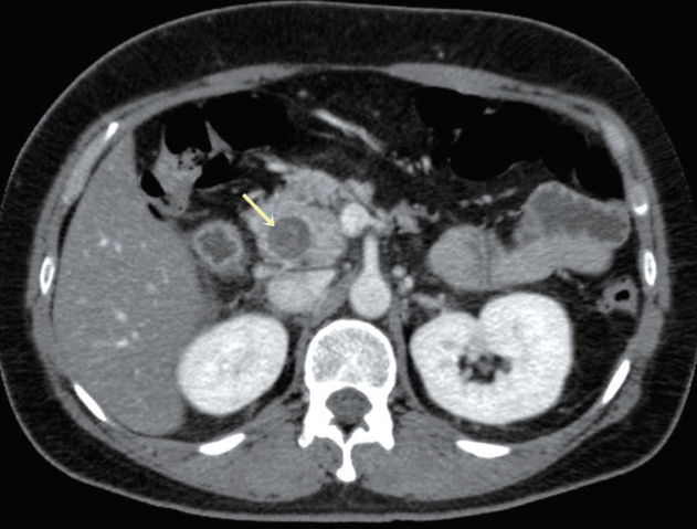
Abdominal computed tomography showed remnant choledochal cyst with a 15 mm sized hyperdense lesion in the pancreatic head portion (arrow).

Magnetic resonance cholangiopancreatography showed remnant choledochal cyst with a signal void lesion in the pancreatic head portion (arrow).
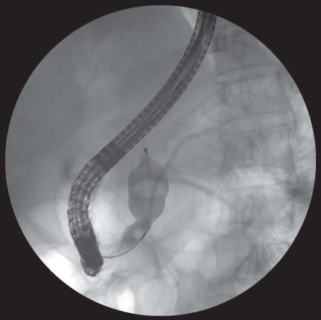
Pancreatic duct stone extraction was done by using a basket under fluoroscopic guidance.
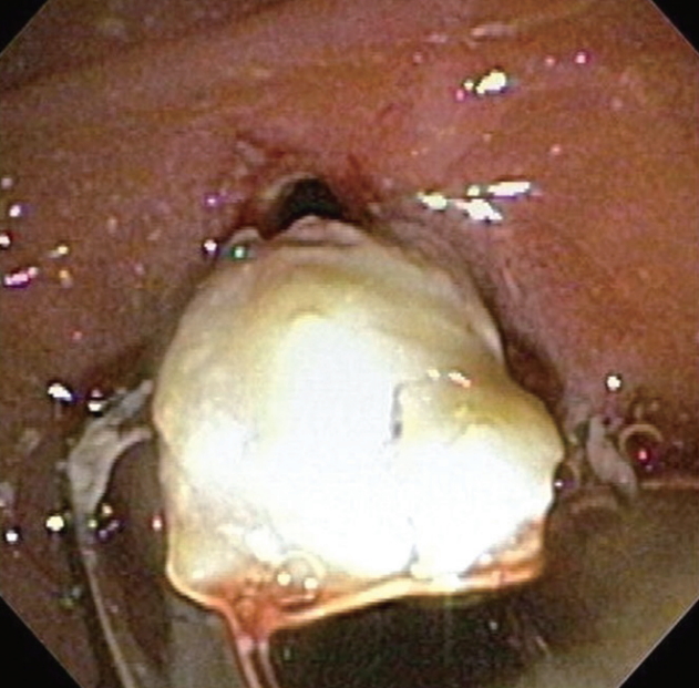
Endoscopic view of the removed pancreatic duct stone. A white, fragile stone was removed after endoscopic sphincterotomy.