담관스텐트의 총담관 천공으로 유발된 혈액담즙증 1예
Delayed Hemobilia Caused by Penetration of Biliary Plastic Stent into Portal Vein
Article information

Abstract
담관 협착 환자에서 내시경적 담관 스텐트 삽입술은 널리 사용되고 있다. 담관 스텐트의 이탈은 삽입 후 발생할 수 있는 합병증 중 하나로 근위측 또는 원위측으로 발생할 수 있으며, 대부분은 무증상이고 이탈한 스텐트의 제거도 성공적으로 이루어진다. 66세의 남자 환자가 국소진행성 췌장선암으로 내원하였다. 종괴가 총담관과 십이지장의 제3부를 침범하여 폐색을 유발하였기 때문에, 환자는 담관 스텐트 삽입술과 이어진 십이지장 스텐트 삽입술을 시행 받고 퇴원하였다. 환자는 3주 후 토혈을 주소로 응급실로 다시 내원하였다. 상부위장관 내시경 및 자기공명영상 검사에서 혈액담즙증의 소견을 보였으나 보존적 치료만으로 환자는 회복되어 퇴원하였다. 출혈의 병소를 찾기 위해 2개월 후 추적한 컴퓨터단층촬영 및 자기공명영상 검사에서 담관 스텐트가 총담관의 내측벽을 뚫고 복강내로 이탈하여 간문맥에 닿은 것이 확인되었다. 이에 담관 스텐트의 이탈과 총담관 천공으로 유발된 혈액담즙증의 1예를 보고한다.
Trans Abstract
Biliary stent migration is one of the late complications of endoscopic retrograde cholangiopancreatography. Most of the biliary stent migration is asymptomatic and successfully managed by stent removal. A 66-year-old man with unresectable pancreatic cancer, common bile duct obstruction, and duodenal third portion obstruction underwent endoscopic plastic biliary stent placement and duodenal uncovered metallic stent placement in two separate sessions. After 3 weeks from the duodenal stenting, he presented with hematemesis. Urgent esophagogastroduodenoscopy and magnetic resonance imaging showed hemobilia. The patient recovered with conservative managements. Cross-sectional imagings done 2 months later demonstrated the penetration of the biliary stent into portal vein. Here, we present a case of delayed hemobilia caused by penetration of biliary plastic stent into the portal vein.
INTRODUCTION
Endoscopic stent placement is a well-established procedure for relieving malignant or benign bile duct stenosis. It has favorable safety profiles. However, there are both early and late onset complications related to endoscopic stent placement. One of the late onset complications is biliary stent migration. This can be either proximal or distal migration. The overall incidence is 3-6% [1].
Proximal stent migration is usually asymptomatic, but it can result in biliary obstruction and cholangitis. In nearly all the cases, proximally migrated stent remains within the biliary tract. The stent migration sticking out of the biliary tract and resulting in hemobilia is very rare. Hence, we report a case of delayed hemobilia caused by penetration of biliary plastic stent into the portal vein.
CASE
A 66-year-old man was referred to Korea University Guro Hospital for the evaluation of pancreatic head mass. He complained of dyspepsia and sense of abdominal discomfort. Past medical history was remarkable for essential hypertension, diabetes mellitus, and a history of thyroid papillary cancer for which the patient had undergone curative resection 6 months ago. Physical examination was unremarkable. Except for an elevated level of serum carbohydrate antigen 19-9 at 2,384 U/mL, complete blood count and biochemical panel were all within normal range. Abdominal computed tomography (CT) showed a 4-cm sized poorly enhanced mass at pancreatic head and uncinate process with invasion of distal common bile duct (CBD) and duodenal third portion (Fig. 1A).
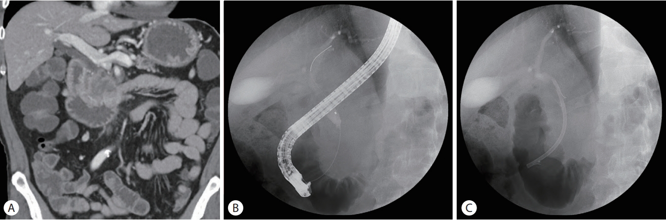
computed tomography and endoscopic retrograde cholangiopancreatography findings. (A) About 4 cm sized poorly enhancing mass was noted in pancreatic head and uncinate process. (B) Cholangiogram showed distal common bile duct obstruction. (C) A 10 Fr×7 cm plastic biliary stent was placed across the biliary obstruction.
According to the multidisciplinary discussion, the neoadjuvant chemotherapy was recommended due to invasion of adjacent major vessels. Following endoscopic ultrasonography guided fine needle aspiration confirmed the histologic diagnosis as pancreatic adenocarcinoma. Liver function tests at that time were as follow: aspartate aminotransferase 21 IU/L, alanine aminotransferase 19 IU/L, alkaline phosphatase 74 IU/L, r-glutamyl transpeptidase 63 IU/L, total bilirubin 1.75 mg/dL, direct bilirubin 0.44 mg/dL. Since liver function tests were anticipated to be worsen, endoscopic retrograde cholangiopancreatography (ERCP) was performed and a 10-Fr 7-cm plastic biliary stent (Cotton-Leung ® Biliary Stent; Cook Medical LLC, Bloomington, IN, USA) was placed without immediate complications. The stent was successfully placed across distal CBD stenosis (Fig. 1B, C). The patient was discharged 2 days after the stent placement with no procedure-related complication and scheduled for the next chemotherapy.
Five weeks later with given two cycles of modified fluorouracil, irinotecan, oxaliplatin and leucovorin (FOLFIRINOX) chemotherapy, the patient visited emergency department with vomiting. CT scan of the abdomen demonstrated worsened duodenal invasion of pancreatic head cancer with luminal obstruction at duodenal third portion (Fig. 2A). Initially, a covered metallic stent placement in the duodenum was tried, but distal migration occurred after a week. Then, endoscopic placement of an uncovered metallic stent (HANAROSTENT ® , M.I. Tech, Pyeongtaek, Korea) was done successfully (Fig. 2B, C). After successful duodenal stent placement, the patient was able to resume oral intake and discharged 3 days later.

Images of duodenal stent placement. (A) Computed tomography of the abdomen showed luminal obstruction at duodenal third portion and proximal luminal dilatation. Previously placed biliary stent was in place. (B) A 2 cm in diameter×8 cm in length, uncovered duodenal metallic stent was inserted under endoscopic guidance. (C) Upper gastrointestinal series showed the locations of the biliary stent and duodenal stent. The arrows indicate the rotated direction of the biliary plastic stent.
After 3 weeks from the duodenal stenting, the patient visited emergency department with hematemesis. The blood pressure of him was 92/64 mmHg and pulse rate was 106 beats per minute. The patient’s hemoglobin was 8.7 g/dL, white blood cell count was 14,500×10³/µL and platelet count was 11,800×10³/µL. Liver function tests were as follow: aspartate aminotransferase 39 IU/L, alanine aminotransferase 18 IU/L, alkaline phosphatase 570 IU/L, r-glutamyl transpeptidase 603 IU/L, total bilirubin 2.75 mg/dL, direct bilirubin 1.67 mg/dL. After transfusion of two units of red blood cells, the hemoglobin level was elevated to 11.3 g/dL.
Urgent esophagogastroduodenoscopy (EGD) showed a large amount of fresh blood was found in duodenal second portion. Initially, it was presumed to be tumor bleeding because no causative lesion for active bleeding was found (Fig. 3A). Subsequent angiography demonstrated no contrast extravasation. Magnetic resonance imaging (MRI) revealed a dark signal intensity in the dependent portion of the common bile duct, which was suggestive of blood clots (Fig. 3B). Also, the axis of the previously placed biliary stent was rotated and the proximal tip was headed towards portal vein. In the follow-up EGD, bleeding from the duodenal second portion has ceased, but there was dark blood clot attached to the major duodenal papilla (Fig. 3C). Fortunately, hemobilia stopped spontaneously and did not recur from then.
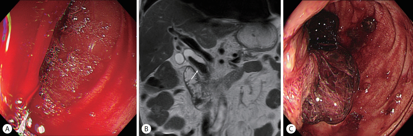
(A) Urgent esophagogastroduodenoscopy showed a large amount of fresh blood in the duodenal second portion. (B) T2-weighted image of magnetic resonance imaging demonstrated a dark signal intensity (arrow) in the dependent portion of the common bile duct, which was suggestive of blood clot. (C) Follow-up esophagogastroduodenoscopy showed a dark-colored blood clot attached to the major duodenal papilla.
Follow-up CT scan and MRI were done 2 months later. Previously placed biliary stent has migrated proximally and its proximal tip penetrated into the medial of the CBD and became attached to the portal vein (Fig. 4). We concluded that the migrated biliary stent penetrated CBD and injured the portal vein, which eventually resulted in hemobilia.
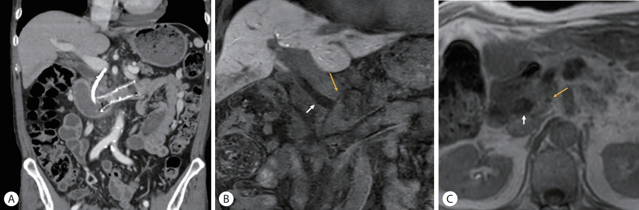
Follow-up cross-sectional images done 2 months later. (A) Abdominal computed tomography. (B) Coronal section of magnetic resonance image. (C) Axial T1-weighted image of the magnetic resonance imaging. All the cross-sectional imaging studies showed the penetration of the biliary stent. The proximal tip of the stent (yellow arrows of B, C) was located outside of the common bile duct wall (white arrows of B, C).
Until now, 5 months after from the event of hemobilia, there has been no recurrence. However, despite of eight cycles of modified FOLFIRINOX chemotherapy administered, the overall response is assessed as stable disease. He is treated with the chemotherapy of same regimen as a maintenance.
DISCUSSION
Hemobilia is potentially a fatal condition and often caused by iatrogenic etiology and bile duct invasion of neoplasm [2]. Hepatic artery is the commonly involved vessel with formation of pseudoaneurysm and arterio-bilio fistula. Bile duct communication with portal vein is very rare, and bleeding from such communication tends to be less severe and often self-limited due to its lower blood pressure gradient [2-4].
Percutaneous transhepatic biliary drainage is highly associated with hemobilia, reported as rate of 2.3-3.0% [2,5,6]. However, hemobilia related with endoscopic biliary stenting is rare, and it is mainly caused by direct injury to the bile duct during procedure. Furthermore, it is very rare that endoscopically inserted plastic stent results in late onset hemobilia. There are a few reported cases, indwelling stent made bile duct erosion and formed a fistula with hepatic artery [7-9]. There is another report where a plastic stent penetrates CBD after 5 weeks from ERCP, and it was suggested that weakened and vulnerable bile duct due to cholecystectomy prior to ERCP, has contributed to stent migration and CBD penetration. The migrated stent was surgically removed and primary repair of CBD penetration was performed [10].
In this case, the biliary plastic stent was rotated in opposite direction against to the CBD angulation probably due to traction of endoscope during duodenal metal stenting procedure. Contrary to distal tip of biliary stent fixed by duodenal angulation and metal stent, its proximal tip could stab the angulated portion of CBD by repetitive endoscopic exam in the period of stenting procedure. As a result, the biliary stent penetrated CBD and erode the portal vein with subsequently resulting in hemobilia. It is thought that the bleeding was self-limited owing to low blood pressure of portal vein and stent occlusion with blood clot.
We did not remove the migrated stent promptly when it was found at out of CBD because the recurrence of hemobilia or bile leakage is worried. There was no evidence of biliary obstruction or cholangitis fortunately, even though it would be impending. Delayed procedure is thought to be beneficial for injured tissue to be healed and fibrotic if the patient is tolerable. It could be a favorable option that replacing plastic stent to a large diameter covered stent to overlay the defect of CBD wall.
Notes
Conflict of Interest
The authors have no conflicts to disclose.