


INTRODUCTION
Ampulla of Vater tumors (AVT) represent a rare tumor entity and bear malignant potential. Radical surgery with pancreaticoduodenectomy (PD) has been considered the standard treatment for this tumor. However, high morbidity and mortality is associated with this procedure and the overall mortality for PD was reported to be 2.1%[1]. Surgical ampullectomy (SA) has existed as an alternative to PD since 1899, but morbidity and mortality of SA was not uncommon[2].
Endoscopic snare papillectomy (ESP) for AVT has been performed instead of SA and outcome of this procedure was similar compared with SA[2]. Hemorrhage, perforation, and pancreatitis are common procedure-related complications of ESP and rare complications such as cholangitis and liver abscess have been reported[3].
Acute acalculous cholecystitis (AAC) stands for cholecystitis without biliary stone and is a potentially devastating entity after surgery. It usually occurs after gastric and colorectal procedures and is rarely reported after ESP[4]. Here, we report a case of AAC after ESP for AVT which was successfully managed with percutaneous cholecystostomy with intravenous antibiotics.
CASE
A 52-year-old man presented for the management of adenoma arising from ampulla of Vater which was found incidentally in routine health check up. He denied any previous medical history and wanted an endoscopic management for the lesion.
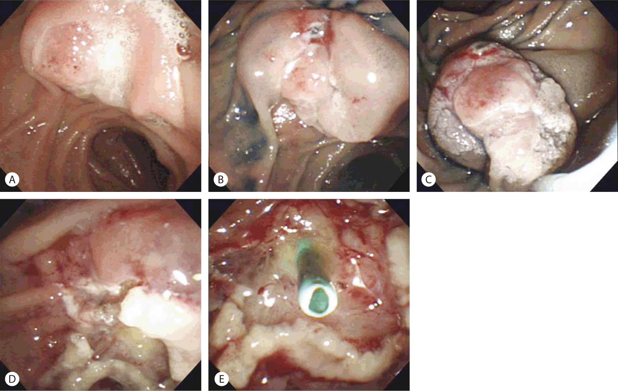
Under conscious sedation with midazolam and meperidine, the lesion was approached with side-viewing scope (Olympus TJF-260V, Tokyo, Japan). After injection of normal saline and epinephrine mixture in the submucosal layer, ESP was performed successfully (Fig. 1) followed by insertion of a pancreatic duct stent (5Fr, 3 cm, Cook Geenen® Pancreatic Stent, Limerick, Ireland). In this step, guide-wire was inserted into the common bile duct and a possibility of guide-wire insertion into cystic duct could not be excluded. Total procedure time was less than 40 minutes. Complications such as major bleeding and/or perforation were not observed. Adenoma with low grade dysplasia was confirmed histopathologically and the margin was clear. Conservative management with intravenous fluid administration and nil per os was maintained.
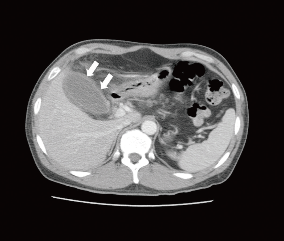
Five days after ESP, the patient complained right upper quadrant pain and the body temperature was elevated to 38.2°C. On laboratory examination, white cell was counted to 20,710/mm3 and amylase was measured to 24 IU/L (reference range 20-100 IU/L). AST and ALT was measured to 17 IU/L (reference range 9-40 IU/L) and 20 IU/L (reference range 0-40 IU/L), respectively. Total bilirubin was 1.7 mg/dL (reference range 0.2-0.4 mg/dL) and direct bilirubin was 0.6 mg/dL (reference range 0-0.3 mg/dL). Abdominal CT showed distended gall bladder with edematous change of the wall without any evidence of biliary stone, which suggested an AAC without any evidence of cholangitis (Fig. 2). Percutaneous cholecystostomy tube was inserted (Fig. 3) and drained under fluoroscopic guidance and intravenous cefoperazone with sulbactam was administered. Ten days after insertion of percutaneous cholecystostomy tube, the patient’s condition improved and laboratory studies returned to normal value. The patient was discharged after removal of percutaneous cholecystostomy tube.
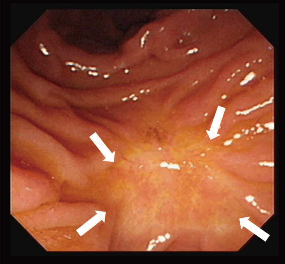
Six months after the procedure, follow-up EGD showed a completely healed ulcer scar at the ampulla of Vater (Fig. 4).
DISCUSSION
Many additional procedures were developed to reduce the complications of EPS. Insertion of a pancreatic duct stent after ESP is one of the best ways to prevent post-procedural pancreatitis[5]. Biliary stenting is less frequently done to reduce the risk of postprocedure cholangitis since this complication is uncommon[6]. Other rare complications such as liver abscess have been reported[3]. Cholecystitis after ESP was reported in a series with giant laterally spreading tumor of ampulla after aggressive endoscopic resection[7]. In another report, cholecystitis was associated with ascending cholangitis[8]. Our case is unique in that it was not associated with ascending cholangitis and aggressive endoscopic resection.
AAC is an acute necroinflammatory disease of the gallbladder with a multifactorial pathogenesis. Gall bladder ischemia is central to the pathogenesis of AAC and is known to be associated with diabetes mellitus, malignant disease, abdominal vasculitis, congestive heart failure, cholesterol embolization, shock, cardiac arrest, non-biliary surgery, and other conditions which could result in a local inflammatory response in the gallbladder wall[9]. Since the patient denied any other systemic diseases, it is plausible that mechanical injury to the cystic duct during guide-wire insertion might have affected the development of AAC in our case.
Cholecystectomy has been considered as a standard therapy for AAC. Percutaneous cholecystostomy is now established as a lifesaving, minimally invasive alternative[10]. Intravenous antibiotics are important adjuncts. In our case, the patient was managed successfully with percutaneous cholecystostomy and intravenous antibiotics. There is no consensus on the optimal time for the tube removal after percutaneous cholecystostomy. But it can be removed when the patients’ symptoms resolves and other laboratory findings and/or imaging studies are in normal range.
In conclusion, AAC even can develop after ESP for small AVT and the endoscopists should perform the procedure meticulously to avoid injury to the cystic duct by guidewire. It can be managed successfully with percutaneous cholecystectomy and intravenous antibiotics.